


Muscles and their attachments in the lower limb play a crucial role in the movements and stability of the body. The lower limb, consisting of the thigh, leg, ankle, and foot, contains some of the largest and strongest muscles in the body. These muscles work together to allow movements such as walking, running, and jumping.
The attachments of these muscles to the bones in the lower limb provide the necessary force and leverage to generate movement. The complex network of muscles and tendons in the lower limb also plays a vital role in maintaining balance and stability while standing or moving.
Understanding the anatomy and function of these structures is essential in the diagnosis and treatment of lower limb injuries and conditions. Moreover, it is important to recognize that the health and fitness of these muscles play a significant role in the overall health and well-being of an individual.
The lower limb muscles are essential for various movements such as walking, running, and jumping. Major muscle groups in the lower limb include the quadriceps, hamstrings, gastrocnemius, soleus, and tibialis anterior, among others.
The quadriceps, located in the front of the thigh, helps to extend the knee joint and is important during daily activities such as climbing stairs. The hamstrings, located in the back of the thigh, flex the knee joint and are necessary for running and jumping. The gastrocnemius and soleus muscles, located in the calf, help with plantar flexion of the ankle joint, enabling us to walk and run.
The tibialis anterior muscle, located in the front of the shin, helps with dorsiflexion of the ankle joint, which is vital for lifting the foot while walking. The proper attachment of these muscles to bones is crucial for normal limb movement. Overall, having knowledge of lower limb muscle anatomy can aid in injury prevention and rehabilitation.
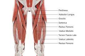
The anterior thigh muscles are an essential group of muscles that are responsible for the movement of the lower limb. The quadriceps femoris muscle group consists of four muscles:
the rectus femoris,
vastus lateralis,
vastus medialis, and
vastus intermedius.
These muscles are located at the front of the thigh and work together to extend the leg at the knee joint. The rectus femoris muscle is unique in that it also flexes the hip joint. The sartorius muscle is another important muscle located at the front of the thigh. It is the longest muscle in the human body and extends from the hip to the medial aspect of the knee joint. Its main function is to flex, abduct, and externally rotate the hip joint and to flex the knee joint.
The posterior thigh muscles, also known as the hamstrings, are a group of three muscles located on the back of the thigh. These muscles include:
the biceps femoris,
semitendinosus, and
semimembranosus.
The hamstrings are responsible for various actions, including hip extension, knee flexion, and internal and external rotation of the leg. They contribute to movements such as walking, running, and jumping.
Additionally, the adductor magnus muscle is responsible for adduction of the thigh, or bringing the leg closer towards the midline of the body. It also plays a role in hip extension and external rotation. The adductor magnus is the largest of the adductor muscles and is often considered a part of the posterior thigh muscles.
The medial thigh muscles are a group of muscles found in the inner thigh that function to adduct or move the leg towards the midline of the body. This group of muscles includes:
the adductor longus,
adductor brevis,
adductor magnus,
gracilis, and
pectineus.
The adductor muscles work together to generate a force that allows the legs to move closer together, while also providing stability to the hip joint during activities such as jumping or running.
Additionally, the obturator externus muscle is located on the lateral surface of the hip and functions to rotate the thigh outward. This muscle is essential for maintaining balance and stability during lower limb movement, and it works in conjunction with other muscles such as the obturator internus and piriformis to keep the pelvis stable during the gait cycle.
The lateral thigh muscles are a group of muscles located on the outside of the thigh. These muscles work together to control movement of the hip and knee joints. The gluteal muscle group, which includes:
the gluteus maximus, medius, and minimus, are the largest muscles in the group and are responsible for hip extension, abduction, and rotation. The tensor fasciae latae muscle is a smaller muscle located in the front of the hip and is responsible for hip flexion and abduction.
It also plays an important role in maintaining stability of the hip joint during weight-bearing activities such as walking and running. Dysfunction of these muscles can lead to a variety of musculoskeletal conditions, including hip pain and instability, knee pain, and lower back pain.
The peroneus longus and brevis muscles are located in the lower leg and are responsible for a range of movements in the foot and ankle. These muscles originate from the fibular head and the lateral condyle of the tibia. The peroneus longus muscle forms a tendon that runs along the outer edge of the foot, while the peroneus brevis tendon runs along the lateral malleolus, the bony prominence on the outside of the ankle.
The peroneus longus muscle addsucts and plantarflexes the foot, while the peroneus brevis muscle assists in plantarflexion and eversion of the foot. These muscles have an essential role in lower limb movement, aiding in stability and propulsion during walking, running, and jumping.
It is essential to understand lower limb muscle anatomy for physical therapy and sports medicine, as proper knowledge of the function and attachments of these muscles can aid in the identification and treatment of muscle injuries and imbalances. Physical therapists can utilize their knowledge of muscle anatomy to design an exercise program that targets specific muscles, supporting the return to function, and reducing the likelihood of reinjury.
Similarly, understanding the anatomy and function of the peroneus longus and brevis muscles can be crucial for sports medicine professionals to help athletes maintain healthy and strong legs and feet.
In conclusion, the peroneus longus and brevis muscles play a critical role in lower limb movement. Understanding their functions, origins, and insertions can be crucial for the prevention and treatment of injuries, making physical therapy more effective and helping athletes perform at their best.
It is important for healthcare professionals specializing in sports medicine and physical therapy to have a solid understanding of lower limb muscle anatomy and function to provide the best care possible to their patients.